Early signs like decreased range of motion may be dismissed as arthritis. For diagnostic purposes, your doctor may recommend undergoing magnetic resonance imaging (MRI).
What Is an ACL Injury?
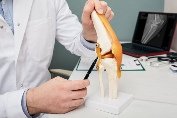
ACL injuries involve a sprain, tear or rupture to the band of tissue connecting the femur to the tibia, or shinbone. While these injuries can happen to anyone, you’re more likely to experience an ACL tear if you engage in activities involving sudden stops, directional changes or jumping and hitting the ground.
The first sign of an ACL injury is a popping sensation in your knee, often followed by visible swelling, decreased range of motion or an unstable joint. Factors increasing your risk for an ACL injury include:
- Being female
- Participating in sports like gymnastics, football, basketball, soccer or skiing
- Poor form or conditioning
- Not wearing proper footwear for a certain activity or the support needed
- Not using effective training equipment
- Playing on a hard, non-responsive surface, like artificial grass
The goal of ACL treatment is to help the area regain its full range of motion and decrease any pain and swelling. Following diagnosis, this may include resting, physical therapy to improve joint strength and stability, or surgery. You might also use a brace or crutches to avoid putting weight and additional stress on your knee.
When you delay getting treatment, you risk developing osteoarthritis in that knee joint.
Diagnosing an ACL Injury with an MRI
For ACL injuries, the diagnostic process starts with a physical exam. Your doctor will look for areas of swelling and tenderness, comparing both knees. You will also be asked to move the joint, so your doctor can compare the range of motion and degree of functionality.
If clear differences are present, your doctor might make a definitive diagnosis or schedule imaging to confirm an ACL injury. MRI is the primary tool, although an X-ray may be ordered to rule out broken bones and an ultrasound to check for the extent of internal injuries.
MRI technology offers the greatest insight, by assessing the bones and soft tissues in detail. It also helps with comparing the state of both knees through visualization of fiber issues in the body. A knee MRI can:
- Help determine the severity of the tear by assessing the ligaments
- Indicate the presence of a partial rupture
- Show if a tibial translocation, fracture or bone bruise is present
However, certain factors can affect the diagnostic process:
- Scarring along the ligament, which may occur if you wait too long to get an MRI
- Blood pooling around the ligament
- Any pre-existing damage or degeneration
Has your doctor recommended a knee MRI to diagnose an ACL injury? Contact Midstate Radiology Associates to make an appointment today.

